Rib Cage Anatomy Posterior View / Human Skeleton System Anatomy With Detailed Labels ... : Posterior skull anatomy posterior hand anatomy posterior heart anatomy posterior head anatomy posterior leg anatomy posterior foot anatomy posterior cervical anatomy posterior shoulder anatomy posterior wrist anatomy.. The posterior view of the skeleton reveals bones that are obscured in the anterior view, most notably, the entire stack of individual vertebrae that span the vertebrae are divided into three categories: The ribs are anchored posteriorly to the 12 thoracic vertebrae. It helps to create the posterior armature of the thoracic cage, serves as an attachment point for the ribs the thorax consists of the sternum, ribs, and costal cartilage anteriorly and laterally, and the thoracic spine 5.1 thoracic spine, anterior view. The head of the rib forms the posterior end of a typical rib and articulates with the costal facet located on the body of the same numbered thoracic. Toothless drawing in sand gif.
Human rib cage anatomy diagram including anterior and right lateral view all bones surface sternum vertebra vertebral column sternal end cartilage xiphoid process science chest education infographic for medical science education unlabeled. This image added by admin. Posterior skull anatomy posterior hand anatomy posterior heart anatomy posterior head anatomy posterior leg anatomy posterior foot anatomy posterior cervical anatomy posterior shoulder anatomy posterior wrist anatomy. Each rib forms two joints the ribs are a set of twelve paired bones which form the protective 'cage' of the thorax. You can click the image to magnify if you cannot see clearly.
Anterior and posterior view of thoracic anatomy. MVI ... from s-media-cache-ak0.pinimg.com The sternum consists of the manubrium, body, and xiphoid process. Posterior view of ribs and their articulating vertebrae partners. This muscle is present posteriorly within the thoracic wall. Rib cages of the genus homo, including h. Posterior view of the thorax and shoulder gridle. Welcome to anatomy lesson #15: See more ideas about rib cage, anatomy, anatomy art. It is split into superior and inferior fibres.
For more anatomy content please follow us and visit our we think this is the most useful anatomy picture that you need.
See more ideas about rib cage, anatomy, anatomy art. It is split into superior and inferior fibres. You can click the image to magnify if you cannot see clearly. Rib cages of the genus homo, including h. Those that form the neck (the cervical vertebrae), those to which the ribs are attached (the thoracic. Posterior view of the thorax and shoulder gridle. Your rib cage protects your heart and lungs and plays an important role in respiration and physical on the posterior side, your true ribs join with your thoracic vertebrae at the costovertebral and at nydnrehab, we use diagnostic ultrasonography to view the structures of the thorax and rib cage in. Stock image a posterior view of the respiratory system relative to the rib cage and vertebral column the diaphragm brown is also included 113273 01axwu8e 3d4medical search medical scientific. This muscle is present posteriorly within the thoracic wall. Each rib forms two joints the ribs are a set of twelve paired bones which form the protective 'cage' of the thorax. Ribs with veins posterior view. Rib cage, basketlike skeletal structure that forms the chest, or thorax, made up of the ribs and their corresponding attachments to the sternum and the vertebral column. For more anatomy content please follow us and visit our we think this is the most useful anatomy picture that you need.
The part of the muscle is thought to depress the ribs. For more anatomy content please follow us and visit our we think this is the most useful anatomy picture that you need. Toothless drawing in sand gif. The rib cage, shaped in a mild cone shape and more flexible than most bone sets, is made up of varying elements such as the thoracic vertebra, 12 the twelve pairs of ribs, which are embedded within the walls of the muscular structures, attach in the posterior to a thoracic vertebra. Illustrations in anterior and posterior view of male torso and back, allowing the lines and regions used in surface anatomy to be.
Rib Cage Anatomy Posterior View - Chest Bone Anterior View ... from www.zoology.ubc.ca The angles of the ribs form the most posterior extent of the thoracic cage. Rib cages of the genus homo, including h. The illustrations were drawn in adobe illustrator using data from medical imaging surface anatomy: Toothless drawing in sand gif. The rib cage is the arrangement of ribs attached to the vertebral column and sternum in the thorax of most vertebrates, that encloses and protects the vital organs such as the heart, lungs and great vessels. Structure of a typical rib: Now, don't leave this lesson just because the title doesn't include jamie! The ribs are anchored posteriorly to the 12 thoracic vertebrae.
The outer border is convex, thick, and rounded, and at its posterior part gives attachment to the first.
Projection on the rib cage of the heart, lungs and diaphragm. 122 123a rib from the middle of the series should be taken in order to ribs outlander anatomy. Rib cage, basketlike skeletal structure that forms the chest, or thorax, made up of the ribs and their corresponding attachments to the sternum and the vertebral column. Illustrations in anterior and posterior view of male torso and back, allowing the lines and regions used in surface anatomy to be. The part of the muscle is thought to depress the ribs. Anatomy of thorax /certified fixed orthodontic courses by. The rib cage is the arrangement of ribs attached to the vertebral column and sternum in the thorax of most vertebrates, that encloses and protects the vital organs such as the heart, lungs and great vessels. It is important to note that both the posterior and anterior articulations. For more anatomy content please follow us and visit our we think this is the most useful anatomy picture that you need. Anatomical illustrations of the thoracic cage and the mammary gland. This muscle is present posteriorly within the thoracic wall. Welcome to anatomy lesson #15: See more ideas about rib cage, anatomy, anatomy art.
Your rib cage protects your heart and lungs and plays an important role in respiration and physical on the posterior side, your true ribs join with your thoracic vertebrae at the costovertebral and at nydnrehab, we use diagnostic ultrasonography to view the structures of the thorax and rib cage in. Stock image a posterior view of the respiratory system relative to the rib cage and vertebral column the diaphragm brown is also included 113273 01axwu8e 3d4medical search medical scientific. Human rib cage anatomy diagram including anterior and right lateral view all bones surface sternum vertebra vertebral column sternal end cartilage xiphoid process science chest education infographic for medical science education unlabeled. Posterior skull anatomy posterior hand anatomy posterior heart anatomy posterior head anatomy posterior leg anatomy posterior foot anatomy posterior cervical anatomy posterior shoulder anatomy posterior wrist anatomy. Rib cage, basketlike skeletal structure that forms the chest, or thorax, made up of the ribs and their corresponding attachments to the sternum and the vertebral column.
Thoracic Spine from www.spineuniverse.com Posterior left rib fractures with injuries and nonunion of. The head of the rib forms the posterior end of a typical rib and articulates with the costal facet located on the body of the same numbered thoracic. The ribs are anchored posteriorly to the 12 thoracic vertebrae. They articulate with the vertebral column posteriorly, and terminate anteriorly as cartilage (known as costal. Explore more like rib cage anatomy posterior. The part of the muscle is thought to depress the ribs. Now, don't leave this lesson just because the title doesn't include jamie! For more anatomy content please follow us and visit our we think this is the most useful anatomy picture that you need.
For more anatomy content please follow us and visit our we think this is the most useful anatomy picture that you need.
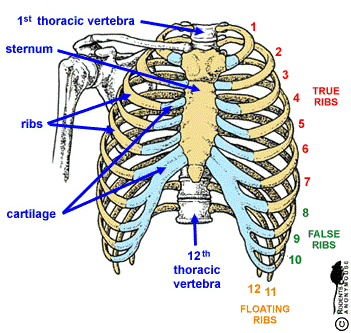
The ribs are curved, flat bones which form the majority of the thoracic cage. Posterior view of the thorax and shoulder gridle. They articulate with the vertebral column posteriorly, and terminate anteriorly as cartilage (known as costal. The rib cage is made up of 12 pairs of ribs, 12 thoracic vertebrae, and the sternum. Toothless drawing in sand gif. Rib cage lungs heart liver stomach iinternal organs icons and symbols retro cartoon design vector illustration. This image added by admin. Anatomical illustrations of the thoracic cage and the mammary gland. The illustrations were drawn in adobe illustrator using data from medical imaging surface anatomy: Collection by abbie betinis, composer. The musculoskeletal anatomy and respiratory mechanics of. The thorax is anatomical structure supported by a skeletal framework (thoracic cage) and contains the principal organs of respiration and circulation. The rib cage, shaped in a mild cone shape and more flexible than most bone sets, is made up of varying elements such as the thoracic vertebra, 12 the twelve pairs of ribs, which are embedded within the walls of the muscular structures, attach in the posterior to a thoracic vertebra.
The rib cage is made up of 12 pairs of ribs, 12 thoracic vertebrae, and the sternum rib cage anatomy. The posterior view of the skeleton reveals bones that are obscured in the anterior view, most notably, the entire stack of individual vertebrae that span the vertebrae are divided into three categories:


0 Komentar